3D dental imaging
Seeing beyond a traditional dental X-ray.
Three-dimensional dental imaging lets us examine teeth, roots, bone and surrounding anatomy from multiple angles when a conventional image does not tell the whole story.
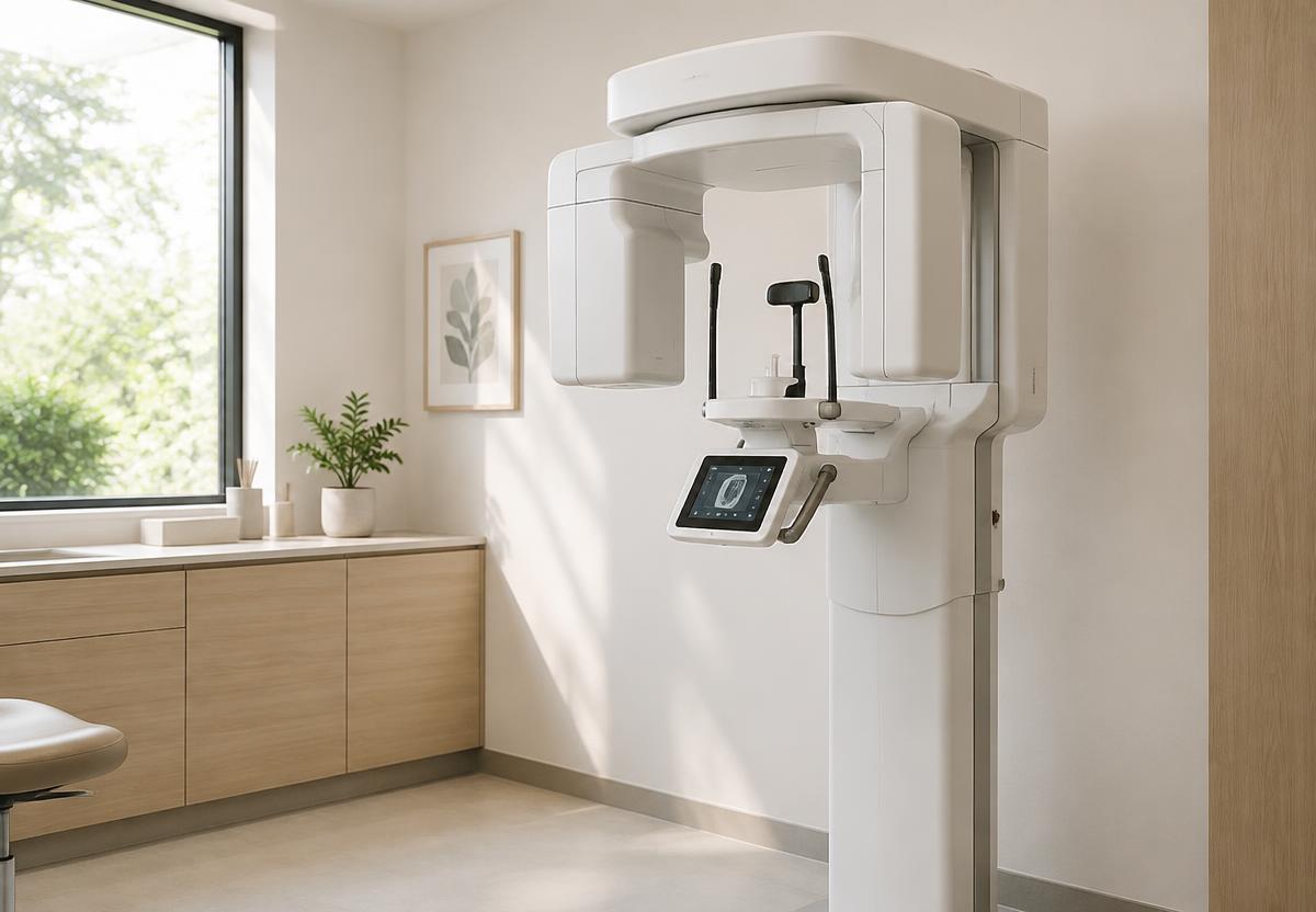
Some dental decisions require more than a two-dimensional image.
Our Carestream Cone Beam Computed Tomography, or CBCT, technology allows us to capture detailed three-dimensional views of your teeth, roots, jawbone and surrounding anatomical structures when that additional information is clinically necessary.
Unlike a conventional dental X-ray, which represents anatomy in two dimensions, CBCT allows us to examine an area from multiple angles and navigate through the anatomy in three dimensions.
That additional information can be invaluable when planning complex treatment or investigating something that cannot be fully understood from conventional imaging alone.
For complex root canal evaluation, three-dimensional imaging is often reviewed alongside treatment carried out under our endodontic microscope.
What it means for you
A three-dimensional view
CBCT allows us to evaluate dental anatomy from multiple planes rather than relying solely on a flat image.
More informed planning
Detailed 3D information can assist in evaluating anatomy for procedures such as implant treatment, endodontic care, oral surgery and other complex cases.
Imaging when it matters
CBCT is used when the additional three-dimensional information is clinically justified, not simply because the technology is available.
Easier explanations
Three-dimensional visualization can help us show you the anatomy we are evaluating and explain the reasoning behind a treatment recommendation.
How it works
In the chair
- The scan itself takes only moments, with you seated or standing still.
- We review the reconstructed images with you where it helps explain a recommendation.
- 3D imaging is requested only when the additional information is clinically justified.
Frequently asked questions
What is a CBCT scan?
CBCT stands for Cone Beam Computed Tomography. It is a dental imaging technique that uses X-rays to produce three-dimensional views of teeth, bone and surrounding anatomical structures.
How is CBCT different from a regular dental X-ray?
Traditional dental X-rays provide two-dimensional images. CBCT creates a three-dimensional dataset that can be examined from different angles and in individual cross-sections.
Do I need a CBCT at every visit?
No. CBCT is used when the additional three-dimensional information is clinically justified. Routine examinations do not automatically require a 3D scan.
What can CBCT help evaluate?
Depending on the case, CBCT can assist with implant planning, root and canal anatomy, impacted teeth, bone structure and other situations where understanding three-dimensional anatomy is important.
More of our technology
Ready when you are.